Oapl Updates

Search
Loading...

5 Tips for Summertime Amputee Skin Care

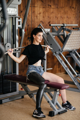
Seasonal changes — spring to summer and summer to fall — present unique challenges for amputee skin care. Skin care plays a pivotal role as you maintain independence and mobility year-round. Whether it’s excess perspiration (summer) or dry skin (winter), most amputees have to make adjustments to their normal skincare routine according to the season. In the summer, hot temperatures and high humidity are the perfect conditions for excess perspiration, heat, and the growth of bacteria and fungus that contribute to a number of skin issues.


Greg Mannino Skiing

We consulted Greg Mannino, a six-time Paralympic gold medalist, prosthetist, and amputee to create a list of summertime skincare tips. With proper skin care, amputees can enjoy the summer sunshine even when the temperatures rise.

1. Cleanse, Cleanse, Cleanse

“Cleanse, cleanse, cleanse,” that’s the number one advice from Mannino. Prostheses create a ready-made breeding ground for heat-related skin issues. The environment inside the prosthesis around the skin is moist and humid with sloughing skin cells. It’s the perfect place for problematic “critters,” as Mannino puts it, like bacteria and fungus to grow and irritate the skin. They’re easy to mistake for heat rash, which can be an issue in and of itself, but good hygiene can prevent painful infections, lesions, ingrown hairs, blisters, and sores.

Most amputees already know that skin hygiene is imperative to their everyday health and success with their prosthesis. But in the summer, skin care needs a new level of attention. Amputees experience more perspiration than average because the body has to compensate for a loss of skin surface area. In addition, prostheses trap body heat and take more physical effort to use, resulting in even more perspiration.

Frequent washing of the residual, liner, and/or sock with a gentle daily cleanser that contains antibacterial and antifungal ingredients keeps the skin and the environment around it as clean as possible. Some amputees can maintain their skin health with one cleansing a day. But others may need two or three cleansing sessions or showers a day followed by regular applications of antibacterial and antifungal products to keep the skin clean.

2. Have a Cleansing Plan When You’re Away From Home

Plan to keep your residual and prosthesis clean while you’re on summer excursions. When you’re at the lake, beach, or on a mountain trail, you may not have quick access to your usual cleanser or a clean water source.

But the heat, moisture, and bacteria build whether you’re close to home or not, and heat and sweat increase bacterial growth. Mannino suggests bringing along a cleanser, liquid-to-powder, and additional socks and liners. In a pinch, a small amount of hand sanitiser can do a quick cleansing until you have access to your cleansing and moisturiser products. The point is that some cleansing, even a small amount, is better than no cleansing at all.

When you’re cleansing in an uncontrolled environment, be careful what the liner, sock, or residual touches after it’s been cleansed and before you put the prosthesis back on. Try to get everything cleaned, dried, and donned without touching surfaces that may harbor bacteria.

3. Change Liners and Socks More Often

“The real key is keeping the socket, liners, and socks clean — the cleaner, the better. The cleaner it is, the less likely you are to have skin breakdown,” says Mannino. You may need a few more extra liners and/or socks in the summer because it’s best to change them frequently. Be sure to cleanse them soon after taking them off so that sweat doesn’t cause odor or make any fabrics harden.

Clean sockets, liners, and socks are also vital because perspiration can cause them to slip and shift when you move. Not only is that uncomfortable, but it can also create sores on the residual and, “chaffing are where bacteria can enter the skin,” warns Mannino.

4. Moisturise Both Day and Night

Heat zaps moisture from the skin, and as the skin gets dry, it can alter the fit of the liner and/or sock. Moisturised skin stays supple and strong, making moisturisers a key part of summertime care. However, make sure the skin is completely dry before donning the prosthesis.

Antibacterial and antifungal moisturisers are a great daytime option because they provide hydration while fighting the growth of unwanted guests. Don’t forget about nighttime moisturization. The skin heals while you sleep, and a night moisturiser aids and promotes that healing.

5. Be Proactive if You Know the Perspiration is Coming

If you know you’re going to work hard, exert yourself, and get sweaty, advanced planning can save your skin and mobility. “In the summer months, you may need to be proactive. If you’re going to sweat more, use a liquid-to-powder preemptively,” suggests Mannino.

A liquid-to-powder product acts as your first defense against excessive perspiration and chaffing. Liquid-to-powder products go on like a regular moisturiser, but they dry to a powder, creating a protective barrier that reduces friction and irritation. These types of products also moisturise to maintain skin integrity. However, for extreme workouts or adventures, when you know you’ll be pushing your physical limits like a race or all-day hike, you can also apply an antiperspirant to your residual the night before.

Conclusion

When all is said and done, “You can prevent many of your own skin issues with good skin care and cleanliness,” says Mannino. Good skin care is part of life for amputees, but a proactive approach during the summer increases your chances of fully participating in your favourite activities. Cleanse, moisturise, and think ahead to make sure your skin stays strong enough for the rigours of life as an amputee.

REQUEST APPOINTMENT

Loading...

Oapl Updates

Search
Loading...

Prosthetic & Orthotic Management of Paediatric Conditions

Orthotic Management of Paediatric Conditions

Orthotists help manage orthopaedic conditions in children with a conservative approach to provide support or alignment correction to reduce pain and optimise their movement.

To clarify, an orthotic is not just something that goes in your shoe! An orthotic is any type of brace that is fitted to any part of your body to support and restore function in the hope to improve mobility, reduce pain and improve overall quality of life both in the short and long term. Below are some of the common reasons why children would need to see an orthotist from a young age.

Neurological Conditions Including Cerebral Palsy

AFO is short for Ankle Foot Orthosis and these types of braces can be used to help position children’s feet who may have weakness or instability in their legs due to Cerebral Palsy and other neurological conditions resulting in foot drop. Traditional AFOs are made of plastic from a cast of someone’s leg which is then used to mould and produce the final device.

Alongside AFOs, there is also the WalkAide available to paediatric patients. The WalkAide uses small electrical stimulations to activate the nerves and muscles in the leg to pick up the foot for children affected with foot drop. Unlike traditional AFOs, the WalkAide is worn on the leg and therefore can be worn barefoot around the home and even on the beach. It works when children walk, run and play, so no matter what activity they are doing they will still be safe and supported.

OAPL are also able to help paediatric clients who require fracture boots, plaster and fibreglass casts, foot orthotics and general bracing for the neck, wrist, knees, and everything in between.

Helmets for Plagiocephaly

Deformational Plagiocephaly often presents as an asymmetrical or uneven head shape. The back of one side of the head will be flattened, often resulting in changes in the forehead and ear alignment. An abnormal head shape caused during the birthing process should begin to correct over time, however if any flat spots are still apparent or new flat areas occur some form of intervention may be required.

In moderate to severe cases of deformational plagiocephaly a cranial remodelling helmet may be recommended. These helmets assist the skull moulding process by removing the pressure over the flat area, allowing the skull to grow into the space provided. Helmets are most effective between 4-12 months of age and results show that helmet therapy can be highly successful in achieving a more symmetrical head shape.

Fortunately, there is no need for a plaster cast when measuring for a remodelling helmet, all measurements are taken with a scanner on a phone and little bubs are free to move as they like! With access to 3D scans and measurements, the family can always track the remodelling process and decide the best course of treatment for their child moving forward.

Developmental Dysplasia of the Hip

Developmental Dysplasia of the Hip (DDH) is the dislocation of the hip due to the shallow and underdeveloped hip socket in some babies. There are various causes of DDH, including family history, breech delivery or a delivery that puts stress on the baby’s hip joints.

DDH is managed with an orthosis that is designed to put the baby’s hips and knees in a bent position to achieve optimal contact between the hip socket and the thigh bone for the hip joint to develop normally.


Prosthetic Management of Paediatric Conditions

Upper limb

Prosthetists don’t just treat lower limb amputations but also treat those with upper limb differences. The majority of upper limb amputee children are the result of congenital limb deficiencies. This can be caused by genetic abnormalities, growth restrictions such as amniotic band syndrome, exposure to viruses in utero and more. A lot of congenital abnormalities don’t always have an explanation as to why and this is particularly the case with upper limb deficiencies.

The sooner a child can begin using a prosthetic arm the better long-term user they will be. Often the first arm will be fitted once the child starts to crawl because they will find it useful when moving around.  These arms would be described as ‘passive’ because they are a hand that doesn’t move. They are shaped to help the child crawl, move objects and carry small items.

Once the child starts to get a bigger and has more cognitive function prosthetists then look at incorporating a hand/hook that has movement. This is normally just the thumb that moves and allows the child to grip and carry objects. The hand/hook moves by the child wearing a harness over their contralateral shoulder and by pulling shoulders forward allows the hand/hook to open and close.

Once a child has mastered using an upper limb prosthesis there are a range of functional attachments for the end of their prosthesis that will give them greater function and ability to try new activities. These might include a tumbler attachment for gymnastics, a guitar pick holder or attachment that allows the child to hold onto the bicycle handle bars.

Lower Limb

As with Upper Limb amputation, the majority of lower limb amputations in children are caused from congenital deficiencies at birth rather than trauma. Often children may be born with deficiencies that effect complete or partial absence of the fibula or tibia. This will result in the lower limb not forming correctly and can affect the length of bones, shape of limb and internal structures of the foot.

In many cases an attempt will be made to keep the limb, however in some cases, this is not possible and the limb will be amputated. If this is the case, it often happens when the child is still a baby.

Once the child has started crawling a cast for a prosthetic limb will be taken. This will be the time it starts to become useful and is often a simple design that is self-suspending.  This will allow the child propulsion when crawling but also let them progress to walking. Design wise, there are a vast range of fun designs that can be incorporated onto children’s legs to help encourage the child to wear them!

Children are amazingly resilient and because they have often grown up using a prosthetic limb they are often excellent walkers. They learn to walk using a prosthesis, grow up using a prosthesis and are able to participate in mostly any activity they choose. Once the child is old enough, prosthetists will endeavour to put them on componentry (Feet/Knees) that will enable them to participate in sport and play with their peers.

REQUEST APPOINTMENT

Loading...

Oapl Updates

Search
Loading...

Managing Heel Pressure and How to Offload

What is Heel Pressure?

Heel pressure can cause serious injuries to the skin behind the heel and if not managed correctly can lead to further ulceration. This happens to people who have poor skin integrity or when people are not mobilising and are experiencing prolonged bed rest. The area behind the heel is prone to breaking down because the heel bone, called the Calcaneus, has a distinct ridge at the back of the heel that makes it prominent. The skin over the area is also thin and does not have much padding, such as muscle or fat that protects the bone from the skin.

Heel pressure occurs when people are laying down and the back of the heel is in contact with the bed. The weight of the leg is passed through the back of the heel where there is minimal padding or resilience to pressure. Movement of the heel on the bed will cause shear stress to the area and the skin can come red and inflamed.

Heel pressure areas are a common occurrence for a range of people. Most often people are experiencing long term bed rest when heel pressures occur. The skin protecting the area is thin and there is little tendon or muscle soft tissue protecting the bone. When people have poor skin integrity and are not mobilising often, they are likely spending a lot of time in bed. When you think about the position they are resting in, you notice their heels are resting right on the bed. Add micro-movements to this time laying down and the skin will experience ongoing shear pressure on the sheets or bed. This can cause pressure areas within hours; It may appear like a red blemish or when you wear a new pair of shoes and develop a heel blister. Or it may be a burst blister and forming into an ulcer.

Symptoms & Causes

As mentioned above, the pressure may first appear as discolouration on the heel, colours to look for are red, purple and blue. Like a bruise, the blood is at the surface of the skin causing the discolouration. It is likely that fluid will develop in the skin like a blister; once the skin breaks, the person is at risk of infection or skin death (necrosis).

There are several known factors that increase a patient’s risk of developing a heel pressure ulcer, including:

  • Inadequate/malnutrition – Poor vitamin intake and absorption from food can lead to thinning of the skin and therefore decreased blood flow.
  • Advancing Age – Elderly people who spend an increased amount of time in bed can lead to shear and pressure in areas of poor skin integrity.
  • Abnormalities of circulation – This may be due to vascular issues where blood flow is limited to the extremities and a lack of red blood cells to an injury will delay healing. This is also common in patients with diabetes.
  • Sensory deficiency – For example, people with diabetes who may experience nerve damage are at increased risk of pressure sores. Nerve damage causing loss of sensation is called Diabetic Neuropathy.
  • Immobility – May be caused by paralysis, stroke or severe illness. Fractures of the bones in the legs may also lead to immobility for a period of time. Common fractures to occur are the neck of the femur (NOF) fractures also known as a broken hip
  • Major surgery – Heart, lung and some orthopaedic surgeries restrict mobility and hence increase time in bed. Long periods of time laying in bed increase chances of pressure sores.
  • Multiple health problems (comorbidities) –  Particularly coronary or respiratory can lead to long term bed rest. Long periods laying in bed increases the chances of pressure sores.
  • Dehydration – Hydration is vital for maintaining skin integrity and wound healing. Adequate fluid intake is necessary to support blood flow to wounded tissues and to prevent additional breakdown of the skin.

Treatment & Prevention

Offloading is described as lifting or pushing an area of high pressure away from the cause of the pressure. To offload is to distribute the load to other areas which are not susceptible to pressure areas. Both the calf and foot can help with the offloading. Heel pressure is redistributed to both the calf, a soft muscle belly which can change shape to fit a supportive device as well as the foot.

Below are some examples of devices available for short term or long-term offloading of the heel.

oapl Heel Cushion

A basic all-rounder ankle, foot and heel relief device which acts as a snug cushion under the foot and ankle. A large hole under the heel allows for the pressure area to float in space and have minimal pressure in the heel region. The device is appropriate for prevention of pressures particularly in the ICU environment. The OAPL Heel cushion comes with a de-rotation wedge which limits rotation of the leg from the hip and keeps the leg in a neutral position. The device also comes with “AFO straps”, elastic straps which can be attached to the heel cushion (via Velcro) in a figure of 8 design, keeping the ankle at 90 degrees, avoiding plantar flexion and contractures occurring.

Heelift

Available in smooth or convoluted foam. The Heelift Suspension Boot is designed to provide an effective solution for prevention and assistive healing of pressure ulcers at the heel. The Heelift delivers positioning and alignment of the lower limb. It is made from latex-free medical grade foam with a friction free tricot (satin) base. This ensures it stays in place on the leg at all times as well as sliding through the bed sheets when required and not sticking. The convoluted design is in place to aid air flow through the device when under the bed sheets. Heelift comes with an extra pad to control hip rotation, foot drop or provide added elevation. One size fits all.

MPO (Multi Podus Orthosis) 

 The RCAI MPO 2000® with Transfer Attachment features a Sky Blue breathable foam liner that wicks moisture away from the skin while maintaining skin integrity. The MPO floats the heel to eliminate pressure or friction on the heel, enhancing blood circulation vital to healing. The dynamic flex action supplies continuous counter force to the plantar surface of the foot assisting in the correction of foot drop, foot and ankle contractures and deformity. The adjustable toe post relieves pressure on the toes and can be positioned to the side in the treatment of malleolus and lateral ulcers. The rotator bar positioned to the side, controls hip and leg rotation, providing functional alignment. The transfer attachment (brown sole) is provided for stand-up transfer and prevents cross-contamination from floor to bed. We do not recommend patients walk in the MPO boot.

REQUEST APPOINTMENT

Loading...

Oapl Updates

Search
Loading...

How Orthotists and Prosthetists are Involved in Treating Diabetic Patients

People often ask how orthotists and prosthetists are involved in treating patients who have diabetes. As a condition that is primarily related to glucose levels in the blood. Having diabetes can lead to an array of health complications if not managed effectively.

Diabetes is a medical condition where the body cannot maintain healthy glucose levels in the blood. When you eat food that contains glucose, your pancreas produces a hormone called ‘Insulin’ that converts glucose from your food into energy. When your body cannot maintain healthy glucose levels, you could have diabetes, a non-curable medical condition that can be serious if not managed correctly.

Type 1 Diabetes

Type 1 diabetes is caused by an autoimmune condition where the body attacks and damages the pancreas and insulin cannot be produced as a result.

Type 2 Diabetes

Type 2 diabetes is caused by lifestyle factors and associated with obesity. It can be caused by the pancreas wearing out by overproducing insulin over a long period of time or if the pancreas has the ability to produce insulin but the muscles and liver prevent the insulin from working. About 1.7 million Australians have diabetes. 10% have type 1 diabetes and 90% of type 2 diabetes. If the glucose is not broken down, it will remain circulating in your blood. When sugar is present in the blood it is called glycemia, and if there is too much glucose in the blood that is unmanaged, it can cause damage to small blood vessels.

Diabetic Foot Ulcer

Complications of Diabetes

Diabetes can lead to damage of nerves and small blood vessels within the body. Damage to nerves that cause loss of sensation in the feet is called peripheral neuropathy. Those with a wound may not feel pain in their feet and therefore may not realise they have a wound under their foot. Poor sensation and blood circulation in the feet increases the risk for sores and skin break down due to poor skin integrity. Being overweight, having poorly fitted shoes or having debris inside your footwear can cause wounds to form. The damage to the body’s arteries and blood vessels lead to poor circulation and can severely affect one’s ability to heal. A normal immune system clears away damaged tissue, builds new skin and fights off infection. If diabetes is unmanaged, the Immune system is weakened and cannot fight off infection and generate new skin.

Management of Complex Diabetes

Diabetic and high-risk foot management involves multi-disciplinary team comprising of podiatrists, endocrinologists, vascular surgery, rehabilitation physicians, clinical psychologists, dieticians, care coordinators and orthotists. This robust team oversees patients with complex diabetic foot  wounds within the inpatient setting or the outpatient setting. The multi-disciplinary team implements evidence-based assessment and management of patients with diabetes related foot problems with the aim of reducing amputation rates, reducing lengths of stay and ensuring cost effective and appropriate use of hospital investigations and resources for this patient group.

Orthotic Management for Diabetics

What is an Orthotist?

An orthotist works with patients who require any form of bracing or support because of issues with the neuromuscular and skeletal systems. Ranging from sports bracing to complex custom supports, an orthotist will assess and treat patients who have physical limitations resulting from llnesses and disabilities. In everyday work, an orthotist will undertake assessments, prescribe, design, fit and adjustments of orthoses. The role of an orthotist within Diabetic and High-risk foot management is an extremely challenging yet important position.

What is an Orthotist’s role in diabetic and high-risk foot management?

Orthotists work with a multidisciplinary team involved in all stages of wound management of diabetic feet to heal wounds and prevent infections through a gold standard of treatment protocols. An orthotist can assess and fit specialised footwear and orthoses to manage active wounds and prevent wounds from recurring after it has healed. Wounds in diabetic patients are slow to heal due to a poor blood supply and reduced immune function. Therefore, there is an increased risk for bacteria to enter the wounds and lead to serious infection.

Pressure, shear or friction can cause break down of skin that has poor supply of oxygen and nutrients. Wounds are often found on the bottom of the feet from the body’s weight. Orthotists aim to reduce pressure under the wound to prevent the wound from getting worst without taking away the patient’s independence and allowing them to continue their day to day activities.

Wounds can be offloaded with a modified post-operative or wound care footwear with felt or a pressure relieving insole. The wound will be relieved by re-directing pressure and forces from weight bearing to a larger area of unaffected skin.

VACOcast Diabetic used in Diabetic Foot Management

Orthotists can also fit a CAM walker to offload wounds on the bottom of the foot for a higher level of offloading. They also have the skill to apply a specialised cast, called a ‘Total Contact Cast’ with the wound protected by felt, layers of cotton and plaster. The cast can re-distribute pressure and weight in the foot and shear and friction during walking.

Even after a wound has healed, a diabetic patient will have a risk of having recurring wounds. An Orthotist is involved in the prescription of a custom insole and footwear that they can wear in the long term. It can be customised to mould the bottom of the patient’s foot, accommodate any deformities and reduce pressure going through areas of the foot that are at risk of foot ulcers. Orthotists work within a high-risk foot team to prevent diabetes-related amputations.

Diabetes and Amputations

Each year in Australia there are more than 4,400 amputations directly related to diabetes and this number is growing. Diabetes causes a decrease in blood flow to the peripheries (feet/hands) and results in reduced sensation and poor healing. Because of these symptoms, diabetic patients are prone to ulcers on their feet/lower limbs and this is the main cause of amputations. If a limb has needed to be amputated there is then far greater risk for the unaffected limb that remains. If the need for amputation is due to poor vascularity, this risk is due to its likely poor circulation, increased load that will be placed through it and the reliance the individual now has on their remaining ‘good’ leg. The chance of having a further amputation almost doubles once the first one has occurred.

Prosthetic Management for Diabetics

Diabetes and caring for your residual limb

After an amputation it is vital to maintain the health of your remaining limb. Due to decreased sensation and the reduction in blood flow, the skin can become more delicate, so its vital to check regularly to ensure there is no redness, rubs, dryness or breakdowns. This can be done by using a hand mirror so a full view of the residuum is possible and should be done at regular periods throughout the day.

What is a prosthetist?

A prosthetist is trained to prescribe, design and fit any prosthetic device used to replace part of a person’s body. They are involved in providing therapy and education around the use of a prosthetic device and how it should serve an individual’s requirements.

What role does a Prosthetist play in diabetic amputations?

After an amputation the individual will need to see a Prosthetist so they can educate the amputee on what they should expect during their recovery and, if appropriate, what the process is for them receiving a new prosthetic limb.

The majority of amputees with diabetes-related amputations will receive some form of prosthesis. It can be used to simply transfer between chairs or walk around the home, go back to work or even begin to play sports. This will all be determined by the amputee’s overall health, ability and goals.

Prosthetic design for patients with Diabetes

An amputee maintaining excellent stump health is vital to a success fitting of a prosthesis. Whilst volume fluctuations are common with all new amputees is can be particularly problematic with diabetic patients. This is because they need to be very diligent in adjusting the fit of their prosthesis with socks to either add or remove space and ensure there is no excess movement. Excess movement is the major cause of skin breakdown and with poor circulation that means delayed healing. This delay in healing will result in the amputee being unable to wear their prosthesis for risk of further skin breakdown and will slow their overall recovery.

Depending on the skin integrity and the level of the amputation the prosthetist will work with the amputee to ensure the most safe and comfortable fit can be achieved. This may be through using gel liners, special socks without seams and a variety of other methods.

REQUEST APPOINTMENT

Loading...

Oapl Updates

Search
Loading...

VACOped – The New Standard for Managing Foot and Ankle Trauma

The VACOped boot by OPED is revolutionising the way we manage foot and ankle fractures and is now considered the gold standard for treatment of Achilles tendon ruptures. Unlike conventional treatments, the VACOped allows the foot and ankle to remain dynamic while still providing the support necessary for a full and quick recovery.

The VACOped utilises VACO12 technology as a main feature of stabilisation. The VACO12 system within the VACOped uses as many as 20 million small polystyrene beads that each make contact with up to 12 other surrounding beads. Energy is transferred along the poly beads but is dispersed and weakened as forces are absorbed from bead to bead.


This limits the chances of movement within the boot due to impact. Like a bean bag, the liner is filled with polystyrene beads that mold around the foot and leg of the wearer. Air is vacuumed out of the liner so that the poly beads contour around the foot and ankle to provide customised protection. This works with a rigid plastic outer shell to make VACOped very cast stable for the wearer.


The VACOped is unique in that it can easily be removed and reapplied. Physicians are able to easily check the fracture site, and physiotherapy can be done at any time. The simplicity with which the VACOped can be removed and reapplied not only means that patients are also able to wash their limb, but the removable liners which cover the vacuum cushion can be easily washed and replaced.

The vacuum technology used in the VACOped’s fracture stabilisation system ensures the boot will re-mold perfectly to the shape of a patient’s limbs, no matter how many times it is removed. The removable sole of the VACO range of boots means they can also be worn at night without soiling bed linen.

The vacuum stabilising system VACOped consists of a modular, honeycomb shaped plastic shell and a vacuum inlay that together offer stability equivalent to a plaster cast. The co-operative patient can put on and remove the VACOped independently. Physical therapy is also possible to preserve dorsiflexion/extension in the ankle joint in the early post-operative phase. Various adapters on the posterior range of motion (ROM) hinge enable restricted dorsiflexion/extension while maintaining lateral stability.


What foot and ankle conditions does the VACOped address and how?

Achilles Tendon Repair

The VACOped was primarily designed and engineered for the treatment of the Achilles tendon rupture. The Range of Motion (ROM) hinge allows the practitioner to set the ankle at different angles of plantar or dorsiflexion and allows for free ROM or a controlled degree of ROM.

Controlled joint movement is an important part of the late rehabilitation phase, allowing the joints and muscles to gradually regain strength. Unlike other conventional treatments which do not allow for any movement, the VACOped ROM function can be set in increments of five degrees.

In the study by Dolphin et.al. Patients who were treated with the VACOped showed better tendon quality post-treatment as well as a decreased rate of re-rupture

Malleolus or Ankle Fractures

The frame design in collaboration with the vacuum cushion allows for cast stable immobilisation in the boot. Unlike other conventional treatments which do not allow for any movement, the VACOped‘s ROM (range of motion) function can be set to allow the patient to walk normally with the boot in situ and ROM at the ankle. This allows the muscles to regain strength and reduces the rate of thrombosis as the calf muscle contracts through gait and moves blood back to the heart.


Unknown Object

How does the VACOped differ from existing technology?

Currently Achilles ruptures are treated in an emergency department with a plaster of paris backslab, the ankle is set in plantar flexion and a crepe bandage wrapped around. In the following days, the patient will attend an orthopaedic clinic where they will be fitted with a CAM walker and a heel wedge. Generally these heel wedges are a stack of 3 or 4 wedges connected to one another but which can be pulled apart. The wedge is positioned under the heel and encourages the ankle into plantar flexion. Because the wedges are bulky and the boot is designed for an ankle in the 90 deg position, the walker and wedge can become ill-fitting and uncomfortable for the patient

The alternate treatment to this is to use a goniometer to measure 30 deg of ankle plantar flexion and the patient has a full plaster cast or synthetic cast applied to the leg. Every two weeks the angle is adjusted by 15 deg until the patient reaches the neutral position. Limitations to this method are: Time to apply the cast, remove and re-apply at least twice. A skilled cast technician is required to set the cast and perform this method. Further limitations include the patient cannot weight bear at all in the cast, particularly when it is set in plantar flexion. This results in the atrophy of the foot and leg muscles.

Conclusion

The VACOped boot represents the modern standard of care for foot, ankle and Achilles injuries. With the ability to provide cast stable immobilisation and a short application time. The VACOped also offers an ankle range of motion function (-15° to +30°). Because the modular boot is designed to move and flex at the ankle it allows true plantar flexion without the ill-fitting nature of the walker and wedge method described above.

VACOped allows for greater freedom of movement than a plaster cast as it can be removed for hygiene reasons or a practitioner wants to check a wound. The total contact nature of the device can significantly reduce recovery times as seen in the study by Honigmann et al.

Clinical Evidence

VACOped Facts & Figures

  • Functional dynamic bracing and functional rehabilitation for Achilles tendon ruptures: a case series Dolphin, P. (Philippa); Bainbridge, K. (Kelly); Mackenney P. (Paul); Dixon, J.(John). 2016
  • Non-surgical treatment of Achilles rupture: Does duration in functional Weight bearing orthosis matter Randeep Aujla  MBChB *, Amit Kumar, Maneesh  Bhatia. 2015
  • The dynamic vacuum orthosis: a functional and economical benefit? Jochen Franke & Sabine Goldhahn & Laurent Audigé&Henry Kohler&Andreas Wentzensen. 2007
  • After treatment of malleolar fractures following  ORIF—functional compared to protected functional in a vacuum-stabilized orthosis: a randomized controlled trial Philipp Honigmann, Sabine Goldhahn, Jan Rosenkranz, Laurent Audigé, Daniel Geissmann, Reto Babst. 2005
  • Cast Immobilization or Vacuum Stabilizing System? U. Stöckle, B. König, A. Tempka, N.P. Südkamp · Trauma and Reconstructive Surgery, Charité, Campus Virchow Klinikum, Humboldt University Berlin. Unfallchirurg. 2000

For more information on the OPED range of boots including the VACOped, VACOcast and VACOpedes, call us on 1300 866 275 or email info@oapl.com.au


REQUEST APPOINTMENT

Loading...

Oapl Updates

Search
Loading...

Understanding Lower Back Pain

The Lower back is a complex structure, made up of 5 lumbar vertebras that support the weight of the upper body. The spine is a crucial part of your body function. The spinal cord that connects the brain to the rest of your body, is protected by the bones in your spine. An injury to any of these structures can result in localised back pain or radiating pain if a nerve that sends signals to the extremity becomes compressed.

Symptoms

Low Back pain, or “Lumbago” can be caused by an injury to a muscle, ligament or joint around the lower spine, called the “lumbar” or “lumbosacral” spine, resulting in localised pain.

Mechanical low back pain can be caused by improper lifting, poor posture, repetitive stress, lack of exercise or fracture to the spine. You can be at higher risk of developing lower back pain from having more pressure or strain on your back by being overweight or pregnant.

Lumbar sprain

Ligaments are the connective tissue that joins bones, joints and cartilage together and keeps the spine stable. The ligaments can be overstretched and tear from improper lifting or lifting something too heavy.

Muscle Spasm

It would be hard for you to distinguish between a ligament or a muscle injury as both can cause significant pain and inflammation in the area. A muscle spasm is an involuntary contraction of the muscle that feels like cramping or tightening. The back muscle would spasm to protect itself or could mean that there is an underlying problem with the spine.

Degenerative disc disease

The ‘disc’ is the layer between the vertebral body that contains a jelly-like substance which acts as a shock absorber in the joint of the spine. It is normal for this layer of disc to become drier and less flexible with age, therefore, your spine will not be able to move freely and can be painful. Your pain can be worst with sitting but can be relieved by standing up, changing positions or lying down.

Ruptured, prolapsed or herniated disc

When the jelly-like substance in the disc bulges out of a tear in the outer casing of the disc, it is called a ‘herniated disc’. In most cases, a herniated disc is undetected as it does not cause symptoms. However, if the bulged disc pushes against a nerve, it will cause a significant amount of pain. Overtime, the herniated portion of the disc can get smaller, therefore symptoms will ease and go away.


Diagnosis

Your doctor may request imaging to find the cause of the pain. An MRI scan can show whether you have an injury soft tissue, spinal discs and nerve roots. Whereas, a CT scan will provide cross-sectional images of your spine.

An X-ray can provide clearer imaging of bone and can be used to rule out any fractures. This will help your doctor decide on how to treat it.


Treatment

Bracing is a conservative approach that provides targeted compression to the muscles and ligaments that are causing pain. It helps relieve muscle tension and pain in your lower back. Bracing can also improve posture to redistribute weight in the spine and to lessen the strain on the muscles and ligaments around the back to provide pain relief during recovery of lower back aggravation.

Hot & Cold Therapy helps alleviate sore and tight muscles around the lower back. Applying heat to an inflamed area will promote blood flow.

Physical Therapy Programs focus on strengthening the core muscle groups in the low back and improve flexibility and posture of the back.

Your doctor may prescribe medications such as pain-relieving medications or non-steroidal anti-inflammatory drugs for acute and chronic low back pain.

oapl Coolfit Cinch

OAPL Cool Fit Cinch

The oapl Coolfit Cinch provides lumbar spine support to help reduce low back pain and support the abdominal region. It features a ventilated mesh support with elastic segment and includes touch tape adjustment for easy application.

Bioskin Back Skin

Bioskin Back skin

Ideal for low back pain due to low back sprains, the Bioskin back skin provides overall compression to your lower back. Can be worn during activities to prevent re-injury by improving back posture. Can be purchased with a foam pad for targeted compression.

Bioskin Vector Back Support

Bioskin Vector

Provides targeted, vectored compression through a dual pulley cinching system through pulling the handles. This will pull the lumbar (lower spine) panel forward into the spine for the targeted compression. The Vector comes with a hot/cold pack that can be inserted into the back section of the brace to provide relief from flare ups from muscular strains and osteoarthritis of the lower back.


oapl have a vast array of experience treating patients with lower back pain. To book an appointment with one of our orthotists please call us on 1300 866 275 or view our clinical locations here.


REQUEST APPOINTMENT

Loading...

Oapl Updates

Search
Loading...

Osteoarthritis of the Knee

Knee Osteoarthritis is a degenerative knee condition where the cartilage of your knee joint gradually wears away, exposing the underlying bone.

Early stages of knee Osteoarthritis (OA) is characterised by the onset of pain and mild inflammation around the knee. Usually pain is more severe in the morning and the knee may feel stiff and take a while to loosen up.


What is Knee Osteoarthritis (OA)?

Osteoarthritis (OA) is the most common form of arthritis and is characterised by the gradual breakdown of the articular cartilage in synovial joints, primarily of the knees, hands, and hips.

What causes Knee Osteoarthritis (OA)?

The body is complex and has processes in place to repair and maintain itself. OA is a result of this process being unable to occur correctly and causes an imbalance in enzymes which leads to the loss of collagen and proteoglycans, two proteins that are essential in the composition of the body’s connective tissue. It can occur spontaneously or be secondary to another condition.

Knee Osteoarthritis (OA)

What are the Risk Factors for Knee Osteoarthritis (OA)?

OA is often associated with joint wear and tear due to aging but can also stem from joint injuries, musculoskeletal abnormalities, genetics, and environmental factors. Obesity exacerbates OA by increasing joint stress and inflammation.

What are the Symptoms of Knee Osteoarthritis (OA)?

Symptoms include pain, swelling, stiffness, and reduced range of motion in the affected joint. Symptoms may worsen in cold weather, upon initiating activity, or after prolonged activity.

How is Knee Osteoarthritis (OA) Evaluated?

Practitioners diagnose OA using radiographic imaging. Key indicators of OA are narrowed joint spaces and bony spur formation which are indicative of cartilage breakdown.

Knee Osteoarthritis (OA) evaluation

What are the Treatment Options for Knee Osteoarthritis (OA)?

While there is no cure, treatment options aim to alleviate pain. Options include pharmaceuticals, physical therapy, bracing, and surgical intervention [1]. Treatment reccomendations will be tailored to the patietnt as well as the severity and location of the OA.

How Does Bracing Help Manage Knee Osteoarthritis (OA)?

Bracing aims to offload or stabilize the affected joint to reduce pain[2,3]. It is often used alongside medication and can be effective for mild to severe OA. Braces will apply three forces, in a ‘push’ or ‘pull’ mechanism, to open the joint space and is primarily beneficial for unilateral OA.

Osteoarthritis knee brace


BRACING OPTIONS

The amount of support provided by the brace is dictated by the severity of the OA and how active a person is. For example, an individual with very severe OA but is not very active, will not require as much support as someone with the same level of OA but is very active. This is because the person who is not as active will not be putting as much weight through the joint which can aggrevate their condition.


Mild support

Recommended for individuals with low OA severity and high activity level or vice-versa.

Action reliever

  • Anatomically shaped knitted fabric provides medical grade compression (~25mmHg)
  • Cross straps deliver an off-loading force that increases when the knee is straight and needs it the most
  • Clinically proven to decrease pain upon exertion and increase walking speed when compared to non-bracing[4].
Airshift

  • Contains an Airbladder system that can be adjusted to the level of unloading needed
  • Re-positionable condylar gel pads focuses the air bladder’s unloading force.
  • Alternating strap patters to prevent rotation of the brace


Moderate support

Ideal for individuals with a moderate to high activity level or OA severity.

UniReliever

  • BOA-dial system for adjustable and precise offloading
  • Cross straps delivers an off-loading force that increases the knee is straight and needs it the most
  • Quick-fit buttons for easy removal and wearing
Aspen OA brace

  • One step closure to for easy removal and wearing
  • Easily adjustable hinge above and below the knee for comfort
  • Comfortable non slip pads


High support

For those with severe OA or a high activity level

Rebel Reliever

  • Loadshifter Technology allows for adjustable corrective forces
  • Clinically found to reduce pain during motion to promote functional ability by decreasing the detrimental forces at the knee during stance[5][6].
  • Available in a range of fun colours


There are many knee brace options availble and finding the most appropriate one can be difficult. Many knee bracing options need to be fit by an expert such as an orthotist or physiotherapist.

See your local oapl orthotist to discuss the most appropriate options.

REQUEST APPOINTMENT

Please contact orthotics@opchealth.com.au for more information.


  1. DeRogatis, M., Anis, H. K., Sodhi, N., Ehiorobo, J. O., Chughtai, M., Bhave, A., & Mont, M. A. (2019). Non-operative treatment options for knee osteoarthritis. Annals of translational medicine, 7(Suppl 7), S245. https://doi.org/10.21037/atm.2019.06.68
  2. Cudejko T, van der Esch M, van den Noort JC, Rijnhart JJ, van der Leeden M, Roorda LD, Lems W, Waddington G, Harlaar J, Dekker J. Decreased pain and improved dynamic knee instability mediate the beneficial effect of wearing a soft knee brace on activity limitations in patients with knee osteoarthritis. Arthritis care & research. 2019 Aug;71(8):1036-43.
  3. Dries, T., VAN DER Windt, J. W., Akkerman, W., Kluijtmans, M., & Janssen, R. P. A. (2022). Effects of a semi-rigid knee brace on mobility and pain in people with knee Osteoarthritis. Journal of rehabilitation medicine. Clinical communications, 5, 2483. https://doi.org/10.2340/jrmcc.v5.2483
  4. Benning, R. Schneider-Nieskens. Superiority of a Knee Relief Orthosis in the Treatment of Knee Osteoarthritis: A Prospective Randomised Controlled Trial. ORTHOPÄDIE TECHNIK. 2017 Aug; 24-30.
  5. Lamberg EM, Streb R, Werner M, Kremenic I, Penna J. The 2-and 8-week effects of decompressive brace use in people with medial compartment knee osteoarthritis. Prosthetics and Orthotics International. 2016 Aug;40(4):447-53.
  6. Thoumie P, Marty M, Avouac B, Pallez A, Vaumousse A, Pipet LP, Monroche A, Graveleau N, Bonnin A, Amor CB, Coudeyre E. Effect of unloading brace treatment on pain and function in patients with symptomatic knee osteoarthritis: the ROTOR randomized clinical trial. Sci Rep. 2018; 8: 10519.
Loading...

Oapl Updates

Search
Loading...

Do you have Achilles Tendonitis?

If you’re experiencing pain and stiffness along your Achilles area then chances are you may have Achilles Tendonitis. Achilles tendonitis is the inflammation of the Achilles tendon above the heel and can be caused by an increase in high impact activities such as running or jumping.

The Achilles tendon joins the calf muscle (Gastrocnemius) to the heel bone (Calcaneus) which is the largest and strongest tendon in the body; however, due to poor blood supply to the tendon, damage takes longer to heal than other soft tissue injuries.

Symptoms & Causes

Achilles tendonitis can present as pain anywhere along the tendon, localised warmth, mild swelling or pink/redness. Pain is intensified when you push up on your toes and lift your heel off the ground. This is due to the tendon contracting and being put under load. Achilles tendonitis is often worsened by:

  • An increase in usual running distance
  • Running uphill frequently which creates a repeated stretch on the Achilles tendon as the ankle is positioned in dorsi flexion (position where the toes point up towards the body)
  • Pronation and over pronation. This is when the foot rolls inwards and the arch of the foot collapses, this position increases the strain on the Achilles and can cause injury
  • Wearing high heels

Achilles tendonitis is a common overuse injury in athletes. The Achilles tendon can be put under stress from overuse of the calves in physical activities such as running and cycling. It is seen in runners who are doing excessive sprinting and speed work but not stretching enough when their calves are tight. Further to this, cyclists whom have their seat position too low are putting their Achilles tendon under stretch due to the foot and ankle being in a dorsiflexed position on the pedals.

Each time the Achilles tendonitis heals, it repairs with a small amount of scar tissue or adhesions. Adhesions will build up over multiple injuries to the Achilles tendon, making the tendon less flexible. Therefore, you should rest and treat the tendonitis as soon as possible. Here are some ways to prevent and treat Achilles tendonitis.


Treatment & Prevention

Stretching
It is important to always stretch the calves, hamstrings and Achilles to maintain strong and flexible leg muscles. One way to stretch the Achilles tendon is to stand with the knees very slightly bent, lean the body forward to reach for the floor. Take deep breaths in and as you exhale allow your body weight to move your torso closer to your knees and your hands will reach further towards your feet. You should feel an obvious stretch behind the knee. This is stretching the hamstrings and the calf muscles.
Another gentle stretch is keeping your feet and heels planted on the floor, shoulder-width apart whilst keeping the torso straight and upright, bend the knees forward. Aim to get your knees bending over the toes or further. This squatting position is stretching the Achilles nicely.

Footwear
If you have Achilles tendonitis, it is best to avoid wearing high heel as wearing heels puts the Achilles tendon in an excessively shortened position. This leads to premature tightness of the tendon and puts them at higher risk of tendon injury when you exercise. Ensure your running shoes are suitable for your foot type. It is important to wear shoes that support the foot and the motions made by the foot whilst running. If you are thinking of increasing your physical activity do it gradually to avoid stress or inflammatory injuries.

Heel Raises
In-shoe heel raises are suitable for pain relief in Achilles tendonitis. A heel raise is a wedge-shaped foam rubber insert, around 6-10mm in height and is placed in the shoe under the heel. Lifting the heel increases the ankle angle and slightly shortens the Achilles tendon to reduce the strain on the tendon. This allows the tendon to heal. You should also limit intense exercise when you have Achilles tendonitis to aid healing, as you risk putting impact and stress through the tendon which can increase the inflammatory response.

Visit www.oaplshop.com.au to see a range of products used in the treatment of Achilles Tendonitis


REQUEST APPOINTMENT

Loading...

Oapl Updates

Search
Loading...

Treating Sever’s Disease

What is Sever’s disease?

Sever’s Disease is a cause of heel pain in children. The disease occurs when the growth plate of the heel is repeatedly injured by excessive forces during adolescence. Typically, sever’s disease is common in physically active growing children. The growth spurt of adolescence commonly occurs anytime between 8-13 for girls and 10-15 for boys and generally, patients will describe a dull ache in the heel, particularly during activity

Common visual symptoms include limping or walking/running with an awkward gait pattern. Parents are usually the first to sight the symptoms, or invariably the child’s coach/teacher. At home, parents can check pain levels when the child rises on to their toes – it will invariably increase. The heel pain is commonly felt on one foot but can be bilateral.


oapl Plantar Fasciitis Sock for Management of Sever’s Disease

Sever’s disease can be treated with a multifactorial approach. Shock absorption around the heel is paramount during both the early and long-term management of the problem. This helps to reduce the accumulative load on the painful region. Our Plantar Fascia support sock will provide appropriate pressure relief under the heel apparatus with our silicone heel cup. This is designed to provide a cushioned and elevated feel under the heel.

Overall, our sock has the capacity to elevate, compress and alleviate heel symptoms particularly in active children who suffer from sever’s disease. This treatment method would be recommended in addition to regularly icing, resting and stretching; in conjunction with wearing comfortable, well fitted shoes as prescribed by your allied health professional.

For more information on the oapl Plantar Fasciitis Sock call us on 1300 866 275 to book an appointment, or view our online shop.

REQUEST APPOINTMENT

Loading...

Oapl Updates

Search
Loading...

The NEW oapl Plantar Fasciitis Sock

OAPL is excited to launch our NEW Plantar Fasciitis Support sock! It has been designed in-house by our team of podiatrists to offer relief of plantar fascial symptoms, heel and arch pain, and ‘first step’ morning pain.

Designed with multizone compression to relieve swelling and discomfort, our new Plantar Fasciitis sock includes an innovative silicone heel cushion for pressure release and advanced cushioning in every step. Providing you with a product that serves as an all day, everyday management solution for a range of heel and foot conditions.

What is Plantar Fasciitis?

Plantar Fasciitis stands for ‘inflammation of the plantar fascia.’ The fascia runs along the bottom of the foot from the heel bone to the toes. It forms part of the arch of the foot and functions as one of our shock absorbing mechanisms. Unfortunately, the cellular makeup of the fascia is not as elastic as muscle tissue and is limited in its ability to elongate or stretch. Functionally, the cellular makeup of the tissue is prone to breakdown given too much traction placed on the fascia (for multiple reasons) leads to microtears, which in turn leads to irritation; inflammation and ultimately pain.

Symptoms wise, Plantar Fasciitis usually causes discomfort and pain in the heel region. Some people make experience arch pain. Both heel and arch pain discomfort are related to Plantar Fasciitis however pain experienced in the heel is far more common than arch pain.

Key features

  • Medical grade compression to reduce swelling and discomfort
  • Anatomic silicone heel insert built into the sock to relieve and elevate the heel
  • Machine washable, breathable fabric
  • Can be worn with or without footwear

Other uses:

  • Chronic sore feet when standing for long periods
  • Elderly patients suffering from fat pad atrophy in the heel
  • Sever’s disease

Sock Care:

The Plantar Fasciitis Support Sock is made from a combination of polyamide, elastane, polyurethane, and cotton. The polyester insert is made from silicone. To ensure correct care of the sock, it is recommended that our sock is hand washed in cool water. The sock should be stored in a cool, dry place away from direct heat or sunlight. It is not suitable for bleaching, ironing, or tumble drying.

Sizing

For more information on our new Plantar Fasciitis Sock, you can view the product on our online shops. Alternatively, feel free to call us on 1300 866 275 or email us at info@oapl.com.au 


REQUEST APPOINTMENT

Loading...